The effect of non-conformal finite element boundaries on electrical monodomain and bidomain simulations
|
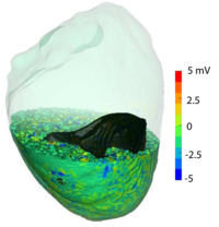
The simulation of electrical activity in the heart, such as normal and abnormal ventricular rhythms and ischemia, utilize computational methods that rely on an underlying geometric model, or polygonal mesh of cardiac tissues and boundaries. Because of the complex shape of many biological structures, it is often difficult to create meshes that conform to the boundaries between distinct regions. The resulting meshes can be non-conformal, i.e., they have element faces that do not align with the surface tangents and the elements represent a smooth surface as a jagged boundary. We hypothesize that these jagged, non-conformal meshes produce local concentrations of current that lead to artifacts large enough to distort the resulting potential fields and generate misleading results. In simulations of acute ischemia, these artifacts can alter the location and severity of the epicardial elevations and depressions, which, in turn, can impact clinical diagnosis. In the case of defibrillation, these artifacts can distort the current density computed through thin structures such as the myocardial wall. |
[DOI/EE link]
@inproceedings{SLFTM10,
  address   = {Belfast, United Kingdom},
  author    = {Darrell J. Swenson and Joshua A. Levine and Zhisong Fu and Jess Tate and Robert S. MacLeod},
  booktitle = {Computing in Cardiology},
  ee        = {https://ieeexplore.ieee.org/abstract/document/5737918/},
  month     = {9},
  pages     = {97--101},
  publisher = {IEEE},
  title     = {The effect of non-conformal finite element boundaries on electrical monodomain and bidomain simulations},
  volume    = {37},
  year      = {2010}
}